Önen, Selin
Loading...

Profile URL
Name Variants
Selin, Önen
Önen,S.
Önen, Selin
O., Selin
Selin, Onen
S.,Önen
O.,Selin
S.,Onen
Onen,S.
Ö.,Selin
Onen, Selin
S., Onen
Önen,S.
Önen, Selin
O., Selin
Selin, Onen
S.,Önen
O.,Selin
S.,Onen
Onen,S.
Ö.,Selin
Onen, Selin
S., Onen
Job Title
Araştırma Görevlisi
Email Address
selin.onen@atilim.edu.tr
Main Affiliation
Basic Sciences
Status
Former Staff
Website
ORCID ID
Scopus Author ID
Turkish CoHE Profile ID
Google Scholar ID
WoS Researcher ID
Sustainable Development Goals
14
LIFE BELOW WATER

0
Research Products
2
ZERO HUNGER

0
Research Products
11
SUSTAINABLE CITIES AND COMMUNITIES

1
Research Products
1
NO POVERTY

0
Research Products
12
RESPONSIBLE CONSUMPTION AND PRODUCTION

0
Research Products
7
AFFORDABLE AND CLEAN ENERGY

0
Research Products
5
GENDER EQUALITY

0
Research Products
3
GOOD HEALTH AND WELL-BEING

4
Research Products
9
INDUSTRY, INNOVATION AND INFRASTRUCTURE

0
Research Products
13
CLIMATE ACTION

0
Research Products
6
CLEAN WATER AND SANITATION

1
Research Products
10
REDUCED INEQUALITIES

0
Research Products
4
QUALITY EDUCATION

0
Research Products
15
LIFE ON LAND

0
Research Products
16
PEACE, JUSTICE AND STRONG INSTITUTIONS

0
Research Products
17
PARTNERSHIPS FOR THE GOALS

0
Research Products
8
DECENT WORK AND ECONOMIC GROWTH

0
Research Products
This researcher does not have a Scopus ID.
This researcher does not have a WoS ID.
Scholarly Output
11
Articles
7
Views / Downloads
1/0
Supervised MSc Theses
0
Supervised PhD Theses
0
WoS Citation Count
90
Scopus Citation Count
96
WoS h-index
5
Scopus h-index
5
Patents
0
Projects
0
WoS Citations per Publication
8.18
Scopus Citations per Publication
8.73
Open Access Source
4
Supervised Theses
0
Google Analytics Visitor Traffic
| Journal | Count |
|---|---|
| Stem Cell Research & Therapy | 2 |
| American Journal of Rhinology & Allergy | 1 |
| Journal of Assisted Reproduction and Genetics | 1 |
| Journal of Hazardous Materials | 1 |
| Progress In Electromagnetics Research Symposium (PIERS) -- AUG 19-23, 2012 -- Moscow, RUSSIA | 1 |
Current Page: 1 / 2
Scopus Quartile Distribution
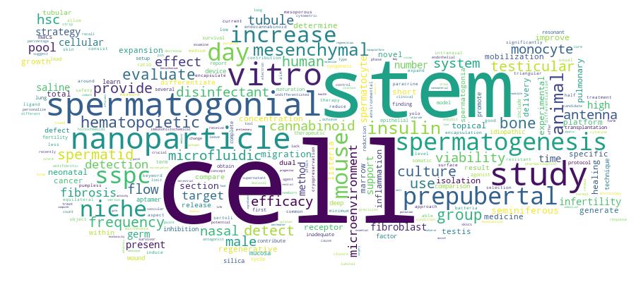
Competency Cloud
11 results
Scholarly Output Search Results
Now showing 1 - 10 of 11
Article Citation - WoS: 2Citation - Scopus: 22-Ag and Bone Marrow-Targeted Pcl Nanoparticles as Nanoplatforms for Hematopoietic Cell Line Mobilization(Bmc, 2024) Kose, Sevil; Varan, Cem; Onen, Selin; Nemutlu, Emirhan; Bilensoy, Erem; Korkusuz, PetekBackgroundThe use of mobilizing agents for hematopoietic stem cell (HSC) transplantation is insufficient for an increasing number of patients. We previously reported lipid made endocannabinoid (eCB) ligands act on the human bone marrow (hBM) HSC migration in vitro, lacking long term stability to be therapeutic candidate. In this study, we hypothesized if a novel 2-AG-loaded polycaprolactone (PCL)-based nanoparticle delivery system that actively targets BM via phosphatidylserine (Ps) can be generated and validated.MethodsPCL nanoparticles were prepared by using the emulsion evaporation method and characterized by Zetasizer and scanning electron microscopy (SEM). The encapsulation efficiency and release profile of 2-AG were determined by liquid chromatography-tandem mass spectrometry (LC-MS/MS). The presence of cannabinoid receptors (CBRs) in HSCs and monocytes was detected by flow cytometry. Cell morphology and viability were assessed using transmission electron microscopy (TEM), SEM, and the WST-1 viability assay. The migration efficacy of the 2-AG and 2-AG-loaded nanoparticle delivery system on HSCs and HPSCs (TF-1a and TF-1) and monocytes (THP-1) was evaluated using a transwell migration assay.ResultsThe 140-225 nm PCL nanoparticles exhibited an increasing polydispersity index (PDI) after the addition of Ps and 2-AG, with a surface charge ranging from - 25 to -50 mV. The nanoparticles released up to 36% of 2-AG within the first 8 h. The 2-AG-Ps-PCL did not affect cellular viability compared to control on days 5 and 10. The HSCs and monocytes expressed CB1R and CB2R and revealed increased migration to media containing 1 mu M 2-AG-Ps-PCL compared to control. The migration rate of the HSCs toward monocytes incubated with 1 mu M 2-AG-Ps-PCL was higher than that of the monocytes of control. The 2-AG-Ps-PCL formulation provided a real time mobilization efficacy at 1 mu M dose and 8 h time window via a specific CBR agonism.ConclusionThe newly generated and validated 2-AG-loaded PCL nanoparticle delivery system can serve as a stable, long lasting, targeted mobilization agent for HSCs and as a candidate therapeutic to be included in HSC transplantation (HSCT) protocols following scale-up in vivo preclinical and subsequent clinical trials.Article Citation - WoS: 4Citation - Scopus: 3Detection of spermatogonial stem/progenitor cells in prepubertal mouse testis with deep learning(Springer/plenum Publishers, 2023) Kahveci, Burak; Onen, Selin; Akal, Fuat; Korkusuz, PetekPurposeRapid and easy detection of spermatogonial stem/progenitor cells (SSPCs) is crucial for clinicians dealing with male infertility caused by prepubertal testicular damage. Deep learning (DL) methods may offer visual tools for tracking SSPCs on testicular strips of prepubertal animal models. The purpose of this study is to detect and count the seminiferous tubules and SSPCs in newborn mouse testis sections using a DL method.MethodsTesticular sections of the C57BL/6-type newborn mice were obtained and enumerated. Odd-numbered sections were stained with hematoxylin and eosin (H&E), and even-numbered sections were immune labeled (IL) with SSPC specific marker, SALL4. Seminiferous tubule and SSPC datasets were created using odd-numbered sections. SALL4-labeled sections were used as positive control. The YOLO object detection model based on DL was used to detect seminiferous tubules and stem cells.ResultsTest scores of the DL model in seminiferous tubules were obtained as 0.98 mAP, 0.93 precision, 0.96 recall, and 0.94 f1-score. The SSPC test scores were obtained as 0.88 mAP, 0.80 precision, 0.93 recall, and 0.82 f1-score.ConclusionSeminiferous tubules and SSPCs on prepubertal testicles were detected with a high sensitivity by preventing human-induced errors. Thus, the first step was taken for a system that automates the detection and counting process of these cells in the infertility clinic.Article Citation - WoS: 24Citation - Scopus: 27Mesenchymal Stem Cells Promote Spermatogonial Stem/Progenitor Cell Pool and Spermatogenesis in Neonatal Mice in Vitro(Nature Portfolio, 2022) Onen, Selin; Kose, Sevil; Yersal, Nilgun; Korkusuz, PetekPrepubertal cancer treatment leads to irreversible infertility in half of the male patients. Current in vitro spermatogenesis protocols and cryopreservation techniques are inadequate to expand spermatogonial stem/progenitor cells (SSPC) from testicles. Bone marrow derived mesenchymal stem cells (BM-MSC) bearing a close resemblance to Sertoli cells, improved spermatogenesis in animal models. We asked if a co-culture setup supported by syngeneic BM-MSC that contributes to the air-liquid interphase (ALI) could lead to survival, expansion and differentiation of SSPCs in vitro. We generated an ALI platform able to provide a real-time cellular paracrine contribution consisting of syngeneic BM-MSCs to neonatal C57BL/6 mice testes. We aimed to evaluate the efficacy of this culture system on SSPC pool expansion and spermatogenesis throughout a complete spermatogenic cycle by measuring the number of total germ cells (GC), the undifferentiated and differentiating spermatogonia, the spermatocytes and the spermatids. Furthermore, we evaluated the testicular cell cycle phases, the tubular and luminal areas using histochemical, immunohistochemical and flow cytometric techniques. Cultures in present of BM-MSCs displayed survival of ID4(+) spermatogonial stem cells (SSC), expansion of SALL4(+) and OCT4(+) SSPCs, VASA(+) total GCs and Ki67(+) proliferative cells at 42 days and an increased number of SCP3(+) spermatocytes and Acrosin(+) spermatids at 28 days. BM-MSCs increased the percentage of mitotic cells within the G2-M phase of the total testicular cell cycle increased for 7 days, preserved the cell viability for 42 days and induced testicular maturation by enlargement of the tubular and luminal area for 42 days in comparison to the control. The percentage of PLZF(+) SSPCs increased within the first 28 days of culture, after which the pool started to get smaller while the number of spermatocytes and spermatids increased simultaneously. Our findings established the efficacy of syngeneic BM-MSCs on the survival and expansion of the SSPC pool and differentiation of spermatogonia to round spermatids during in vitro culture of prepubertal mice testes for 42 days. This method may be helpful in providing alternative cures for male fertility by supporting in vitro differentiated spermatids that can be used for round spermatid injection (ROSI) to female oocyte in animal models. These findings can be further exploited for personalized cellular therapy strategies to cure male infertility of prepubertal cancer survivors in clinics.Article Citation - WoS: 1Citation - Scopus: 2Cannabinoid Receptor Ligands Modulate Fibrosis and Inflammation in Idiopathic Pulmonary Fibrosis: a Preliminary Study(Tubitak Scientific & Technological Research Council Turkey, 2024) Kose, Sevil; Onen, Selin; Gizer, Merve; Boduroglu, Esin; Gonullu, Ugur; Korkusuz, PetekBackground/aim: No specific pharmacological treatment regimen for idiopathic pulmonary fibrosis (IPF) exists. Therefore, new antiinflammatory therapeutic strategies are needed. Cannabinoids (CBs), known for their inflammation-modulating and antifibrotic effects, may be potential medication candidates for treating IPF. We aim to evaluate the inflammation-modulating and antifibrotic effects of CB receptor (CBR) agonists and antagonists in lipopolysaccharide-stimulated normal human lung fibroblast, epithelial cells, IPF fibroblast cells, and monocytes. Materials and methods: We detected CBRs in normal human lung fibroblasts (LL24) and IPF fibroblast cells (LL29), epithelial cells (A549) and monocytes (THP-1) by flow cytometry. We determined TGF-(31, IL-8, and TNF-alpha inflammatory cytokines in the LL24, LL29, A549, and THP-1 cell culture supernatants on days 1 and 5 by ELISA. We evaluated the cell viability in LL24, LL29, and A549 cells on days 1, 3, and 5 spectrophotometrically and detected collagen Type I (ColI) production in the LL24 and LL29 cell culture supernatants on days 1, 3, and 5 by ELISA. Results: LL24, LL29, A549, and THP-1 cells exhibited CB1 (CB1R) and CB2 (CB2R) receptors. CB1R and CB2R agonists WIN55,2122 and JWH015 inhibited fibroblastic and epithelial cell proliferation on day 5. TGF-(31 and TNF-alpha release increased, while IL-8 release decreased in LL24, LL29, A549, and THP-1 cells in response to the administration of WIN55,212-2 and JWH015 at a 10-2 mM concentration. CB1R and CB2R antagonists AM251 and AM630 did not block agonistic responses, suggesting a nonclassical CBRmediated pathway. CB2R agonist JWH015 decreased ColI expression in IPF lung fibroblasts LL29 on day 3. Conclusion: These results suggest that CB signaling regulates the progression of pulmonary inflammation and fibrosis via CBR activation. This may offer a potential pharmacological tool for developing antifibrosis therapies.Book Part Citation - WoS: 15Citation - Scopus: 15Comparison of Hematopoietic and Spermatogonial Stem Cell Niches From the Regenerative Medicine Aspect(Springer international Publishing Ag, 2018) Kose, Sevil; Yersal, Nilgun; Onen, Selin; Korkusuz, PetekRecent advances require a dual evaluation of germ and somatic stem cell niches with a regenerative medicine perspective. For a better point of view of the niche concept, it is needed to compare the microenvironments of those niches in respect to several components. The cellular environment of spermatogonial stem cells' niche consists of Sertoli cells, Leydig cells, vascular endothelial cells, epididymal fat cells, peritubular myoid cells while hematopoietic stem cells have mesenchymal stem cells, osteoblasts, osteoclasts, megacaryocytes, macrophages, vascular endothelial cells, pericytes and adipocytes in their microenvironment. Not only those cells', but also the effect of the other factors such as hormones, growth factors, chemokines, cytokines, extracellular matrix components, biomechanical forces (like shear stress, tension or compression) and physical environmental elements such as temperature, oxygen level and pH will be clarified during the chapter. Because it is known that the microenvironment has an important role in the stem cell homeostasis and disease conditions, it is crucial to understand the details of the microenvironment and to be able to compare the niche concepts of the different types of stem cells from each other, for the regenerative interventions. Indeed, the purpose of this chapter is to point out the usage of niche engineering within the further studies in the regenerative medicine field. Decellularized, synthetic or non-synthetic scaffolds may help to mimic the stem cell niche. However, the shared or different characteristics of germ and somatic stem cell microenvironments are necessary to constitute a proper niche model. When considered from this aspect, it is possible to produce some strategies on the personalized medicine by using those artificial models of stem cell microenvironment.Book Part Citation - WoS: 7Citation - Scopus: 11Magnetic-Based Cell Isolation Technique for the Selection of Stem Cells(Humana Press inc, 2019) Korkusuz, Petek; Kose, Sevil; Yersal, Nilgun; Onen, SelinMagnetic-activated cell sorting (MACS) is the technology that is recently used as a magnetic-based cell isolation/purification technique. This technique enables the isolation and selection of germ, hematopoietic, and somatic stem cells including skin stem cells (SkSCs). Here, we have tried to describe the isolation of stem cells by MACS using CD34 antigen for SkSCs, again CD34 for hematopoietic stem cells (HSCs) and Thy-1 for spermatogonial stem cells (SpSCs). MACS allowed the isolation of CD34+, CD34+, and Thy-1+ human SkSCs, HSCs, and SpSCs with minimum 98% purity.Conference Object Citation - WoS: 4Dual-Frequency, Two Shorting Pin-Loaded Equilateral Triangular Patch Antennas(Electromagnetics Acad, 2012) Can, Sultan; Kapusuz, K. Yavuz; Aydin, ElifThis study presents the resonant frequencies of several antennas, which are dual-frequency antennas. These antennas possess two shorting pins to form dual frequency. Among several methods to provide dual operation, two shorting pins are used to form dual frequency equilateral triangular antennas since there are limited studies on this issue and higher frequency ratios are achievable. The present study has been done with this in mind in order to contribute to related literature on such antennas. Here, the parameters that affect the resonant frequencies of the antenna are also evaluated. The thickness, side length, and shorting pin positions are changed numerous times, and the resonant frequencies are examined according to the change of these parameters. Finally, the frequency ratios are determined for each variation, and the results are compared. The proposed antennas in present study achieved a frequency ratio around 5.7, which is significantly higher from the ones presented in the literature.Article Citation - WoS: 16Citation - Scopus: 19A Pumpless Monolayer Microfluidic Device Based on Mesenchymal Stem Cell-Conditioned Medium Promotes Neonatal Mouse in Vitro Spermatogenesis(Bmc, 2023) Onen, Selin; Atik, Ali Can; Gizer, Merve; Kose, Sevil; Yaman, Onder; Kulah, Haluk; Korkusuz, PetekBackgroundChildhood cancer treatment-induced gonadotoxicity causes permanent infertility/sub-infertility in nearly half of males. The current clinical and experimental approaches are limited to cryopreservation of prepubertal testicular strips and in vitro spermatogenesis which are inadequate to achieve the expanded spermatogonial stem/progenitor cells and spermatogenesis in vitro. Recently, we reported the supportive effect of bone marrow-derived mesenchymal cell co-culture which is inadequate after 14 days of culture in static conditions in prepubertal mouse testis due to lack of microvascular flow and diffusion. Therefore, we generated a novel, pumpless, single polydimethylsiloxane-layered testis-on-chip platform providing a continuous and stabilized microfluidic flow and real-time cellular paracrine contribution of allogeneic bone marrow-derived mesenchymal stem cells.MethodsWe aimed to evaluate the efficacy of this new setup in terms of self-renewal of stem/progenitor cells, spermatogenesis and structural and functional maturation of seminiferous tubules in vitro by measuring the number of undifferentiated and differentiating spermatogonia, spermatocytes, spermatids and tubular growth by histochemical, immunohistochemical, flow cytometric and chromatographic techniques.ResultsBone marrow-derived mesenchymal stem cell-based testis-on-chip platform supported the maintenance of SALL4(+) and PLZF(+) spermatogonial stem/progenitor cells, for 42 days. The new setup improved in vitro spermatogenesis in terms of c-Kit(+) differentiating spermatogonia, VASA(+) total germ cells, the meiotic cells including spermatocytes and spermatids and testicular maturation by increasing testosterone concentration and improved tubular growth for 42 days in comparison with hanging drop and non-mesenchymal stem cell control.ConclusionsFuture fertility preservation for male pediatric cancer survivors depends on the protection/expansion of spermatogonial stem/progenitor cell pool and induction of in vitro spermatogenesis. Our findings demonstrate that a novel bone marrow-derived mesenchymal stem cell-based microfluidic testis-on-chip device supporting the maintenance of stem cells and spermatogenesis in prepubertal mice in vitro. This new, cell therapy-based microfluidic platform may contribute to a safe, precision-based cell and tissue banking protocols for prepubertal fertility restoration in future.Article Citation - WoS: 1Citation - Scopus: 1Topical Intranasal Insulin Enhances Healing of Nasal Mucosa: an Experimental Animal Study(Sage Publications inc, 2023) Kulekci, Cagri; Ozer, Serdar; Onen, Selin; Korkusuz, Petek; Yilmaz, TanerObjective Aim of this study was to evaluate the effect of topical intranasal insulin on healing of nasal mucosa in a rat model. Methods Forty-eight Wistar rats, weighing between 250 and 300 g and aged 10-12 weeks were used and randomized into two equal groups. 1.9 mm curette was introduced through the left nostril and 1.9 mm mucosa from the left nasal septum was curetted. Postoperatively, animals in the control group received 1 mL of physiologic saline, 3 times a day in a nasal irrigation fashion. Animals in the experimental group received 1 mL of 5 IU/mL regular insulin in saline solution. Subjects were sacrificed after 5, 10, and 15 days and macroscopic and histomorphometric evaluations were performed. Results There were no mucosal synechiae and septal perforation macroscopically. Histological examination revealed that the defect size reduction was 21% in the saline group versus 56% in the insulin group on the fifth day (p = 0.006). There was 62% defect reduction in the saline group versus 79% in the insulin group on the 10th day (p = 0.034). On the 15th day, only 67% of saline group animals had complete defect closure, whereas 100% of animals treated with insulin had complete closure (92% vs 100% mucosal defect reduction, p = 0.036). Both edema and inflammation were less in the insulin group on 15th day (p = 0.006; p = 0.023, respectively). Conclusion The results from this study support the safety and efficacy of topical insulin on wound healing in the literature. This study could guide further experimental studies that examine human sinonasal wound healing.Article Citation - WoS: 16Citation - Scopus: 16Targeted mesoporous silica nanoparticles for improved inhibition of disinfectant resistant Listeria monocytogenes and lower environmental pollution(Elsevier, 2021) Sudagidan, Mert; Yildiz, Gulsah; Onen, Selin; Al, Rabia; Temiz, S. Sevval Nur; Yurt, Mediha Nur Zafer; Ozalp, Veli C.Benzalkonium chloride (BAC) is a common ingredient of disinfectants used for industrial, medical, food safety and domestic applications. It is a common pollutant detected in surface and wastewaters to induce adverse effects on Human health as well as aquatic and terrestrial life forms. Since disinfectant use is essential in combatting against microorganisms, the best approach to reduce ecotoxicity level is to restrict BAC use. We report here that encapsulation of BAC in mesoporous silica nanoparticles can provide an efficient strategy for inhibition of mi-crobial activity with lower than usual concentrations of disinfectants. As a proof-of-concept, Listeria mono-cytogenes was evaluated for minimum inhibitory concentration (MIC) of nanomaterial encapsulated BAC. Aptamer molecular gate structures provided a specific targeting of the disinfectant to Listeria cells, leading to high BAC concentrations around bacterial cells, but significantly reduced amounts in total. This strategy allowed to inhibition of BAC resistant Listeria strains with 8 times less the usual disinfectant dose. BAC encapsulated and aptamer functionalized silica nanoparticles (AptBACNP) effectively killed only target bacteria L. monocytogenes, but not the non-target cells, Staphylococcus aureus or Escherichia coli. AptBACNP was not cytotoxic to Human cells as determined by in vitro viability assays.
