Yığman, Zeynep
Loading...
Profile URL
Name Variants
Yigman, Zeynep Z.,Yığman Y.,Zeynep Zeynep, Yigman Zeynep, Yığman Yigman,Z. Yığman,Z. Z.,Yigman Y., Zeynep Z., Yigman Yığman, Zeynep
Job Title
Doktor Öğretim Üyesi
Email Address
zeynep.yigman@atilim.edu.tr
Main Affiliation
Basic Sciences
Status
Former Staff
Website
ORCID ID
Scopus Author ID
Turkish CoHE Profile ID
Google Scholar ID
WoS Researcher ID
No research topics data found.
Sustainable Development Goals
 1
1NO POVERTY
0
Research Products
 2
2ZERO HUNGER
0
Research Products
 3
3GOOD HEALTH AND WELL-BEING
4
Research Products
 4
4QUALITY EDUCATION
0
Research Products
 5
5GENDER EQUALITY
0
Research Products
 6
6CLEAN WATER AND SANITATION
0
Research Products
 7
7AFFORDABLE AND CLEAN ENERGY
0
Research Products
 8
8DECENT WORK AND ECONOMIC GROWTH
0
Research Products
 9
9INDUSTRY, INNOVATION AND INFRASTRUCTURE
0
Research Products
 10
10REDUCED INEQUALITIES
0
Research Products
 11
11SUSTAINABLE CITIES AND COMMUNITIES
0
Research Products
 12
12RESPONSIBLE CONSUMPTION AND PRODUCTION
0
Research Products
 13
13CLIMATE ACTION
0
Research Products
 14
14LIFE BELOW WATER
2
Research Products
 15
15LIFE ON LAND
0
Research Products
 16
16PEACE, JUSTICE AND STRONG INSTITUTIONS
0
Research Products
 17
17PARTNERSHIPS FOR THE GOALS
0
Research Products
This researcher does not have a Scopus ID.
This researcher does not have a WoS ID.
No records found in other affiliations.
Scholarly Output
12
Articles
12
Views / Downloads
50/15
Supervised MSc Theses
0
Supervised PhD Theses
0
WoS Citation Count
56
Scopus Citation Count
49
Patents
0
Projects
0
WoS Citations per Publication
4.67
Scopus Citations per Publication
4.08
Open Access Source
9
Supervised Theses
0
| Journal | Count |
|---|---|
| Drug Design, Development and Therapy | 2 |
| Journal of Updates in Cardiovascular Medicine | 2 |
| Medicina | 2 |
| Medicina (Lithuania) | 1 |
| Scientific Reports | 1 |
Current Page: 1 / 2
Scopus Quartile Distribution
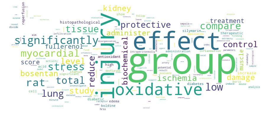
Competency Cloud
12 results
Scholarly Output Search Results
Now showing 1 - 10 of 12
Article Potential Protective Effects of Boldine in Rat With an Experimental Myocardial Ischemia-Reperfusion Model(2025-03-25) Küçük, Ayşegül; Dursun, Alı Dogan; Arslan, Mustafa; Sezen, Şaban Cem; Yıldırım, Alperen Kutay; Özer, Abdullah; Yığman, ZeynepObjectives: Myocardial ischemia-reperfusion injury (MIRI) remains a major challenge in cardiovascular medicine due to its complex pathophysiology involving oxidative stress, inflammation, and cellular dysfunction. Boldine, a potent natural alkaloid with antioxidant and anti-inflammatory properties, has demonstrated protective effects in various pathological conditions. However, its potential cardioprotective effects in MIRI remain largely unexplored. This study aims to evaluate the protective effects of Boldine in a rat model of MIRI by assessing oxidative stress markers, histopathological changes, and inflammatory responses. Materials and Methods: Male Albino Wistar rats were randomly assigned to four groups: Control, Boldine, myocardial ischemia-reperfusion (MIR), and myocardial ischemia-reperfusion + Boldine (MIR+B). Myocardial ischemia was induced by ligating the left anterior descending coronary artery for 30 minutes, followed by 120 minutes of reperfusion. Boldine (50 mg/kg) was administered intraperitoneally at the onset of reperfusion. Cardiac tissue samples were collected for histopathological evaluation and biochemical analysis, including total antioxidant status (TAS), total oxidant status (TOS), and Oxidative Stress index (OSI). Results: Histopathological analysis revealed significant myocardial disorganization and inflammation in the MIR group compared to controls (p=0.05). Boldine treatment significantly reduced inflammation and myocardial disorganization in the MIR+B group (p<0.05), suggesting a protective effect. Biochemical analysis showed a marked decrease in TAS levels and an increase in TOS and OSI in the MIR group (p<0.001). However, Boldine administration significantly restored TAS levels and reduced TOS and OSI in the MIR+B group (p< 0.001), indicating attenuation of oxidative stress. Conclusion: Boldine exhibits significant cardioprotective effects in a rat model of MIRI by reducing oxidative stress, mitigating myocardial disorganization, and alleviating inflammation. These findings suggest that Boldine may serve as a therapeutic agent in ischemic heart disease. Further research is warranted to elucidate its precise mechanisms of action and potential clinical applications.Article Effectiveness of Boric Acid in Sepsis in Rats With Cecal Perforation(Springer Nature, 2025-11-28) Kurtipek, Ali Can; Dursun, Ali Dogan; Yigman, Zeynep; Ozdemir, Cagri; Kucuk, Aysegul; Gonullu, Ugur; Arslan, MustafaIntroduction and AimSepsis is a systemic inflammatory response that develops in the host against microorganisms, which results in end-organ damage. Boric acid (BA) has been shown to have immune modulatory effects in vitro and in animal studies. The aim of the study is to investigate the effects of high dose BA on lung and kidney tissues in rats with sepsis induced by the CLP method.Method28 rats were randomly divided into four groups: Group C (control group), Group BA, Group CLP (cecal ligation and puncture), and Group CLP + BA. Cecum was ligated below the ileocecal valve and punctured. BA was administered to the treatment groups at an intraperitoneal dose of 200 mg/kg, and at the end of 24 h, lung and kidney tissue samples were collected and evaluated for biochemical and histopathological parameters.ResultsHistopathologically, in kidney tissue, CLP + BA group showed significantly less peritubular capillary dilatation and brush border loss in the proximal tubule epithelium compared to the CLP group. In lung tissue, CLP + BA group had significantly less alveolar wall thickening compared to the CLP group. Biochemical analyses indicated that BA administration reduced oxidative stress in both renal and lung tissues.ConclusionWe found that intraperitoneal administration of high dose boric acid partially ameliorated the tissue damage in rats subjected to CLP induced sepsis. Further studies are needed regarding the dosage and application at different time points.Article Citation - WoS: 9Ozone Administration Reduces Myocardial Ischemia Reperfusion Injury in Streptozotocin-Induced Diabetes Mellitus Rat Model(Baycinar Medical Publishing, 2024-11-01) Gulcan, M.B.; Demirtas, H.; Ozer, A.; Yıgman, Z.; Dursun, A.D.; Arslan, M.; Oktar, G.L.Objective: This study aimed to investigate the effects of ozone therapy on myocardial ischemia/reperfusion injury in a diabetic rat model. Methods: The experimental study included 38 male Wistar Albino rats weighing between 200 and 250 g. The rats were randomly assigned to five groups. The sham group included six rats, while the other groups had eight rats each. The other groups were the diabetic ozone group, the diabetic group, the diabetic ischemia/reperfusion group (DIR), and the diabetic ischemia/reperfusion ozone group (DIRO). A total of 32 rats received 65 mg/kg streptozotocin, and a week after the administration, diabetes was confirmed by measuring blood sugar. The rats were fed ad libitum for 40 days to reveal macrovascular complications of diabetes. Malondialdehyde, catalase, superoxide dismutase, paraoxonase-1, total oxidative status, total antioxidant status, and oxidative stress index were assessed. A TUNEL (terminal deoxynucleotidyl transferase dUTP nick end labeling) assay was employed to assess apoptosis. Results: Histologic and biochemical assessments showed the benefits of ozone in myocardial ischemia/reperfusion injury in diabetic rats. The DIRO group was found to be superior to the DIR group. Conclusion: Ozone has cardioprotective effects in streptozotocin-induced diabetic rats through its antioxidant properties against oxidative stress. The study is unique in terms of ozone’s protective effects in diabetic rats against myocardial ischemia/reperfusion injury. However, further studies are needed to confirm our findings. © (2024), (Baycinar Medical Publishing). All rights reserved.Article Citation - WoS: 12Citation - Scopus: 13Protective Effects of Hydrogen Rich Saline Solution in Rats With Experimental Myocardial Ischemia Reperfusion Injury(Cell Press, 2023-12) Koksal, Zeynep; Kurtipek, Omer; Arslan, Mustafa; Dursun, Ali Dogan; Yigman, Zeynep; Ozer, AbdullahAim: The aim of our study is to show whether the administration of hydrogen-rich saline solution (HRSS) intraperitoneally before left main coronary artery (LAD) ischemia protects the myocardium against ischemia-reperfusion (IR) injury.Materials and methods: After ethics committee approval, 24 Wistar Albino rats were divided into 4 groups, 6 rats in each group. For experimental IR, myocardial ischemia was performed by LAD ligation. Left thoracotomy was performed without ischemia in the Control group (Group C). Left thoracotomy was performed without myocardial ischemia to the rats in the HRSS group, and HRSS was given intraperitoneally (ip) at a rate of 10 ml/kg throughout the procedure. In the MIRHRSS group, a single dose of 10 ml/kg HRSS was administered 5 min before reperfusion. Histopathological and biochemical parameters were compared in myocardial tissue samples taken at the end of the reperfusion period.Results: When the groups were compared among themselves in terms of TOS and TAS levels, there was a significant difference between the groups (p = 0.006, p = 0.002). The severity of cardiomyocyte degeneration was significantly greater in MIR group than that in the control and HRSS groups (p = 0.002 and p = 0.001, respectively), as well as severity score of cardiomyocyte degeneration was higher in MIR-HRSS group compared with HRSS group (p = 0.035).Conclusion: Our study shows that HRSS is protective in IR injury, with the application of HRSS 5 min before reperfusion, interstitial edema severity, subendocardial haemorrhage are reduced, and oxidant status parameters are increased, while antioxidant status parameters are decreased. We believe that when it is supported by other studies, the protective effects of HRSS on IR damage will be shown in detail and its indications will be expanded.Article Citation - WoS: 7Citation - Scopus: 6The Effect of Cerium Oxide (ceo<sub>2</Sub>) on Ischemia-Reperfusion Injury in Skeletal Muscle in Mice With Streptozocin-Induced Diabetes(Mdpi, 2024-04-30) Ozer, Abdullah; Sengel, Necmiye; Kucuk, Ayseguel; Yigman, Zeynep; Ozdemir, Cagri; Kilic, Yigit; Arslan, MustafaObjective: Lower extremity ischemia-reperfusion injury (IRI) may occur with trauma-related vascular injury and various vascular diseases, during the use of a tourniquet, in temporary clamping of the aorta in aortic surgery, or following acute or bilateral acute femoral artery occlusion. Mitochondrial dysfunction and increased basal oxidative stress in diabetes may cause an increase in the effects of increased reactive oxygen species (ROS) and mitochondrial dysfunction due to IRI. It is of great importance to examine therapeutic approaches that can minimize the effects of IRI, especially for patient groups under chronic oxidative stress such as DM. Cerium oxide (CeO2) nanoparticles mimic antioxidant enzymes and act as a catalyst that scavenges ROS. In this study, it was aimed to investigate whether CeO2 has protective effects on skeletal muscles in lower extremity IRI in mice with streptozocin-induced diabetes. Methods: A total of 38 Swiss albino mice were divided into six groups as follows: control group (group C, n = 6), diabetes group (group D, n = 8), diabetes-CeO2 (group DCO, n = 8), diabetes-ischemia/reperfusion (group DIR, n = 8), and diabetes-ischemia/reperfusion-CeO2 (group DIRCO, n = 8). The DCO and DIRCO groups were given doses of CeO2 of 0.5 mg/kg intraperitoneally 30 min before the IR procedure. A 120 min ischemia-120 min reperfusion period with 100% O-2 was performed. At the end of the reperfusion period, muscle tissues were removed for histopathological and biochemical examinations. Results: Total antioxidant status (TAS) levels were found to be significantly lower in group DIR compared with group D (p = 0.047 and p = 0.022, respectively). In group DIRCO, total oxidant status (TOS) levels were found to be significantly higher than in group DIR (p < 0.001). The oxidative stress index (OSI) was found to be significantly lower in group DIR compared with group DCO (p < 0.001). Paraoxanase (PON) enzyme activity was found to be significantly increased in group DIR compared with group DCO (p < 0.001). The disorganization and degeneration score for muscle cells, inflammatory cell infiltration score, and total injury score in group DIRCO were found to be significantly lower than in group DIR (p = 0.002, p = 0.034, and p = 0.001, respectively). Conclusions: Our results confirm that CeO2, with its antioxidative properties, reduces skeletal muscle damage in lower extremity IRI in diabetic mice.Article Citation - WoS: 12Citation - Scopus: 17Therapeutic Efficacy of Boric Acid Treatment on Brain Tissue and Cognitive Functions in Rats With Experimental Alzheimer's Disease(Dove Medical Press Ltd, 2023-05) Ozdemir, Cagri; Arslan, Mustafa; Kucuk, Aysegul; Yigman, Zeynep; Dursun, Ali DoganIntroduction: Oxidative stress has an important role in the pathophysiology of Alzheimer's disease (AD), the most common type of dementia. Boric acid (BA) contributes significantly to the protection of the brain by reducing lipid peroxidation and supporting antioxidant defense. We aimed to evaluate the therapeutic potential of BA treatment in AD rats. Materials and Methods: Four groups were formed as Control (C), Alzheimer's (A), Alzheimer's + Boric acid (ABA), Boric acid (BA). Intracerebroventricular injection of Streptozotocin (STZ) was preferred to create an AD. After 4 weeks, BA was applied 3 times every other day. The Radial Arm Maze Test (RAMT) was used to evaluate memory and learning abilities. Biochemical and histopathological evaluations were made in the hippocampus. Results: Initial RAMT inlet/outlet (I/O) numbers were similar. Two weeks after STZ injection, I/O numbers decreased in group A and ABA compared to group C and BA (p<0.05). After the second BA application, I/O numbers increased in the ABA group compared to the A group (p<0.05). In group A, PON-1, TOS and OSI levels were higher and TAS levels were lower than in groups BA and C. After BA treatment, PON-1 and OSI levels were lower in the ABA group than in the A group (p<0.05). Although there was an increase in TAS value and a decrease in TOS, this did not make a statistical difference. The thickness of the pyramidal cell in CA1 and the granular cell layers in the dentate gyrus, and the number of intact and degenerated neurons in the pyramidal cell layer were similar between the groups. Discussion: Significant improvement in learning and memory abilities after BA application is promising for AD. Conclusion: These results show that BA application positively affects learning and memory abilities, and reduces oxidative stress. More extensive studies are required to evaluate histopathological efficacy.Article Effects of Pomegranate Seed Oil on Lower Extremity Ischemia-Reperfusion Damage: Insights into Oxidative Stress, Inflammation, and Cell Death(MDPI, 2025-01-24) Bozok, Ummu Gulsen; Ergorun, Aydan Iremnur; Kucuk, Aysegul; Yigman, Zeynep; Dursun, Ali Dogan; Arslan, MustafaAim: This study sought to clarify the therapeutic benefits and mechanisms of action of pomegranate seed oil (PSO) in instances of ischemia–reperfusion (IR) damage in the lower extremities. Materials and Methods: The sample size was determined, then 32 rats were randomly allocated to four groups: Control (C), ischemia–reperfusion (IR), low-dose PSO (IR + LD, 0.15 mL/kg), and high-dose PSO (IR + HD, 0.30 mL/kg). The ischemia model in the IR group was established by occluding the infrarenal aorta for 120 min. Prior to reperfusion, PSO was delivered to the IR + LD and IR + HD groups at doses of 0.15 mL/kg and 0.30 mL/kg, respectively, followed by a 120 min reperfusion period. Subsequently, blood and tissue specimens were obtained. Statistical investigation was executed utilizing Statistical Package for the Social Sciences version 20.0 (SPSS, IBM Corp., Armonk, NY, USA). Results: Biochemical tests revealed significant variations in total antioxidant level (TAS), total oxidant level (TOS), and the oxidative stress index (OSI) across the groups (p < 0.0001). The IR group had elevated TOS and OSI levels, whereas PSO therapy resulted in a reduction in these values (p < 0.05). As opposed to the IR group, TASs were higher in the PSO-treated groups. Histopathological analysis demonstrated muscle fiber degeneration, interstitial edema, and the infiltration of cells associated with inflammation in the IR group, with analogous results noted in the PSO treatment groups. Immunohistochemical analysis revealed that the expressions of Tumor Necrosis Factor-alpha (TNF-α), Nuclear Factor kappa B (NF-κB), cytochrome C (CYT C), and caspase 3 (CASP3) were elevated in the IR group, while PSO treatment diminished these markers and attenuated inflammation and apoptosis (p < 0.05). The findings demonstrate that PSO has a dose-dependent impact on IR injury. Discussion: This research indicates that PSO has significant protective benefits against IR injury in the lower extremities. PSO mitigated tissue damage and maintained mitochondrial integrity by addressing oxidative stress, inflammation, and apoptotic pathways. Particularly, high-dose PSO yielded more substantial enhancements in these processes and exhibited outcomes most comparable to the control group in biochemical, histological, and immunohistochemical investigations. These findings underscore the potential of PSO as an efficacious natural treatment agent for IR injury. Nevertheless, additional research is required to articulate this definitively.Article Bosentan Reduces Myocardial Ischemia-Reperfusion Injury in Rats(2025-10-01) Küçük, Ayşegül; Dursun, Alı Dogan; Arslan, Mustafa; Özer, Abdullah; Demirtas, Huseyin; Gulcan, Mehmet Burak; Yığman, ZeynepObjectives: This study aimed to investigate the cardioprotective effects of bosentan, an endothelin receptor antagonist, against myocardial ischemia-reperfusion injury (MIRI) in rats. Materials and Methods: Twenty-four adult Wistar-Albino rats were randomly divided into four groups: control, bosentan only, myocardial ischemia-reperfusion (MIR), and MIR-bosentan (MIR-B). Ischemia was induced by ligation of the left anterior descending coronary artery for 30 minutes, followed by 90 minutes of reperfusion. Bosentan was administered intraperitoneally at 30 mg/kg during ischemia in the MIR-B group. Histopathological evaluation assessed neutrophil infiltration, cardiomyocyte damage, tissue edema, and hemorrhage, while biochemical analyses measured total oxidant status (TOS), total antioxidant status (TAS), oxidative stress index (OSI), and paraoxonase-1 (PON-1) activity in myocardial tissue. Results: The MIR group showed significantly increased histopathological injury scores, including neutrophil infiltration, cardiomyocyte damage, edema, and hemorrhage, compared to control and bosentan-only groups (p<0.001). Bosentan treatment significantly reduced these injury scores in the MIR-B group compared to the MIR group (p<0.05). Biochemically, the MIR group exhibited elevated TOS and OSI levels and reduced TAS and PON-1 activity, indicating oxidative stress. Bosentan administration significantly improved these parameters by lowering TOS and OSI levels, and by increasing TAS and PON-1 activity compared to the MIR group (p<0.05). Conclusion: In conclusion, bosentan demonstrated significant protective effects against MIRI by attenuating histological damage and oxidative stress in rat myocardium. These findings suggest that endothelin receptor antagonism with bosentan may offer a promising therapeutic approach to reduce myocardial injury following ischemia-reperfusion events such as those occurring during coronary artery bypass grafting. Further studies are needed to explore its clinical potential.Article Citation - WoS: 1Citation - Scopus: 1Organ-Protective Effects of Fullerenol and Desflurane in a Rat Model of Ischemia–Reperfusion Injury(Nature Portfolio, 2025-11-17) Kip, Gulay; Koksal, Zeynep; Yigman, Zeynep; Kucuk, Aysegul; Arslan, Mustafa; Akarca Dizakar, Saadet Ozen; Sivgin, VolkanTo investigate the protective effects of fullerenol applied before ischemia induction and desflurane anesthesia applied during ischemia-reperfusion (IR) induction in the lungs and kidneys of a lower-extremity IR injury rat model. After receiving ethical approval, we randomly divided 30 rats into five groups: sham (S), IR, IR with 100 mg/kg fullerenol (IR-FUL), IR with 6.7% desflurane (IR-DES), IR with 100 mg/kg fullerenol and 6.7% desflurane (IR-FUL-DES). Fullerenol was administered 30 min before the IR procedure in the IR-FUL and IR-FUL-DES groups, and desflurane was administered during the IR procedure in the IR-DES and IR-FUL-DES groups. During the procedure, an atraumatic microvascular clamp was placed in the aorta for 120 min. The clamp was then removed to achieve reperfusion for 120 min. Finally, at the end of reperfusion, we evaluated the extracted lung and kidney tissue samples and assessed them biochemically and histopathologically. The lung damage scores of the IR-FUL, IR-DES, and IR-FUL-DES groups were significantly lower than those of the IR group (p < .0001, p = .002, and p < .0001, respectively). The renal tubule injury scores of the IR, IR-FUL, IR-DES, and IR-FUL-DES groups were significantly higher than those of the S group (p < .0001). By contrast, the renal tubule injury scores of the IR-FUL and IR-FUL-DES groups were significantly lower than those of the IR group (p < .0001 and p = .001, respectively). Moreover, kidney intercellular adhesion molecule 1 (ICAM1) expression was significantly lower in all the treatment groups, particularly the IR-FUL group, than in the IR group, and lung ICAM1 expression was significantly lower in the IR-FUL and IR-FUL-DES groups than in the other treatment groups. In the lung and kidney tissues, thiobarbituric acid reactive substance levels, catalase activity, glutathione-S-transferase activity, and arylesterase activity were relatively high in the treatment groups. The application of fullerenol before and after desflurane anesthesia during IR has protective effects on rat lungs and kidneys. In particular, histopathology confirmed that the application of fullerenol 30 min before IR induction and desflurane anesthesia during IR induction reduced oxidative stress and alleviated IR-related damage in the lungs and kidneys. These findings may have important translational relevance, suggesting potential perioperative strategies for protecting organs from ischemia-reperfusion injury in clinical settings.Article Citation - WoS: 5Citation - Scopus: 8Protective Effects of Bosentan Via Endothelin Receptor Antagonism in Experimental Ischemia-Reperfusion Injury in the Lower Limb of Rats(Dove Medical Press Ltd, 2025-03) Demirtas, Hueseyin; Oezer, Abdullah; Guelcan, Mehmet Burak; Yigman, Zeynep; Kuecuek, Ayseguel; Tekin, Esra; Arslan, Mustafa; Özer, Abdullah; Gülcan, Mehmet Burak; Küçük, AyşegülObjective: This study aimed to evaluate the protective effects of bosentan, a dual endothelin receptor antagonist, against skeletal muscle ischemia-reperfusion injury (IRI) in rats. Methods: A total of 24 male Wistar Albino rats were divided into four groups: control (C, n=6), bosentan-treated (B, n=6), ischemiareperfusion (IR, n=6), and bosentan plus ischemia-reperfusion (B+IR, n=6). Bosentan (10 mg/kg) was administered 30 minutes prior to reperfusion. In the IR and B+IR groups, ischemia was induced using vascular bulldog clamps for 45 minutes, followed by 120 minutes of reperfusion. Results: Histological and biochemical assessments revealed significant differences among the groups. The disorganization and degeneration scores of the muscle cells in the B+IR group were significantly lower than those in the IR group (P = 0.001). The degree of interstitial edema in the IR group was markedly more severe than in the C and B groups (all P < 0.001), while the interstitial edema score in the B+IR group was significantly lower than that in the IR group (P < 0.001). The total muscle injury scores were markedly reduced in the B+IR group compared to the IR group (P < 0.001). Biochemically, TAS levels were significantly higher in the B+IR group compared to the IR group (1.03 f 0.18 vs 0.59 f 0.10 mmol/L, P = 0.016). Conversely, TOS (1.97 f 0.39 vs 2.86 f 0.43 IU/mg, P < 0.001) and OSI levels (P < 0.001) were significantly lower in the B+IR group. Additionally, paraoxonase (PON-1) enzyme activity was significantly reduced in the B+IR group compared to the IR group (P < 0.001). These findings suggest that bosentan exerts its protective effects by antagonizing endothelin-1 receptors, thereby mitigating vasoconstriction, oxidative stress, and inflammation. The observed reductions in muscle cell disorganization, interstitial edema, hemorrhage, neutrophil infiltration and oxidative stress markers underscore bosentan's potential as a therapeutic agent for managing ischemia-reperfusion injury. Conclusion: Bosentan demonstrates significant protective effects against skeletal muscle IRI by reducing oxidative stress and inflammation through endothelin receptor antagonism. These findings underscore bosentan's potential as a therapeutic agent for mitigating ischemia-reperfusion injury in vascular surgeries and managing critical limb ischemia in clinical settings. Further research is warranted to explore the long-term effects of bosentan on muscle recovery and systemic health following ischemia-reperfusion injury.