Bektur Aykanat, Nuriye Ezgi
Loading...

Profile URL
Name Variants
Bektur Aykanat, Nuriye Ezgi
B.A.Nuriye Ezgi
B. A. Nuriye Ezgi
N., Bektur Aykanat
Bektur Aykanat,N.E.
Bektur Aykanat N.
N.E.Bektur Aykanat
B.,Nuriye Ezgi
B., Nuriye Ezgi
N.,Bektur Aykanat
Bektur Aykanat,Nuriye Ezgi
Nuriye Ezgi, Bektur Aykanat
N. E. Bektur Aykanat
Nuriye Ezgi Bektur Aykanat
Aykanat, Nuriye Ezgi Bektur
Bektur, Ezgi
Job Title
Doçent Doktor
Email Address
ezgi.bekturaykanat@atilim.edu.tr
Main Affiliation
Basic Sciences
Status
Website
ORCID ID
Scopus Author ID
Turkish CoHE Profile ID
Google Scholar ID
WoS Researcher ID
Sustainable Development Goals
3
GOOD HEALTH AND WELL-BEING

5
Research Products
5
GENDER EQUALITY

1
Research Products
16
PEACE, JUSTICE AND STRONG INSTITUTIONS

1
Research Products

This researcher does not have a Scopus ID.

Documents
0
Citations
0

Scholarly Output
9
Articles
8
Views / Downloads
38/0
Supervised MSc Theses
0
Supervised PhD Theses
0
WoS Citation Count
21
Scopus Citation Count
29
WoS h-index
3
Scopus h-index
4
Patents
0
Projects
0
WoS Citations per Publication
2.33
Scopus Citations per Publication
3.22
Open Access Source
7
Supervised Theses
0
Google Analytics Visitor Traffic
| Journal | Count |
|---|---|
| Canadian Journal of Physiology and Pharmacology | 2 |
| Turkish Journal of Medical Sciences | 2 |
| Uludağ Üniversitesi Tıp Fakültesi Dergisi | 2 |
| Annali Italiani di Chirurgia | 1 |
| Osmangazi Tıp Dergisi | 1 |
Current Page: 1 / 2
Scopus Quartile Distribution
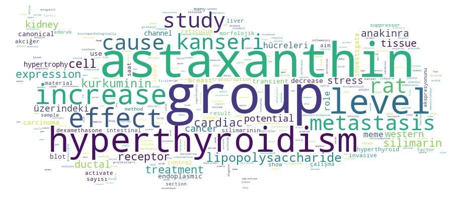
Competency Cloud

9 results
Scholarly Output Search Results
Now showing 1 - 9 of 9
Article Citation - WoS: 3Citation - Scopus: 5Investigation of the Effect of Hyperthyroidism on Endoplasmic Reticulum Stress and Transient Receptor Potential Canonical 1 Channel in the Kidney(Tubitak Scientific & Technological Research Council Turkey, 2021) Aykanat, Nuriye Ezgi Bektur; Şahin, Erhan; Kaçar, Sedat; Bağcı, Rıdvan; Karakaya, Şerife; Dönmez, Dilek Burukoğlu; Şahintürk, VarolBackground/aim: Hyperthyroidism is associated with results in increased glomerular filtration rate as well as increased renin-angiotensin-aldosterone activation. The disturbance of Ca2+ homeostasis in the endoplasmic reticulum (ER) is associated with many diseases, including diabetic nephropathy and hyperthyroidism. Transient receptor potential canonical 1 (TRPC1) channel is the first cloned TRPC family protein. Although it is expressed in many places in the kidney, its function is uncertain. TRPC1 is involved in regulating Ca2+ homeostasis, and its upregulation increases ER Ca2+ level, activates the unfolded protein response, which leads to cellular damage in the kidney. This study investigated the role of TRPC1 in the kidneys of hyperthyroid rats in terms of ER stress markers that are glucose-regulated protein 78 (GRP78), activating transcription factor 6 (ATF6), (protein kinase R (PKR)-like endoplasmic reticulum kinase) (PERK), Inositol-requiring enzyme 1 (IRE1). Materials and methods: Twenty male rats were assigned into control and hyperthyroid groups (n = 10). Hyperthyroidism was induced by adding 12 mg/L thyroxine into the drinking water of rats for 4 weeks. The serum-free T3 and T4 (fT3, fT4), TSH, blood urea nitrogen (BUN), and creatinine levels were measured. The histochemical analysis of kidney sections for morphological changes and also immunohistochemical and western blot analysis of kidney sections were performed for GRP78, ATF6, PERK, IRE1, TRPC1 antibodies. Results: TSH, BUN, and creatinine levels decreased while fT3 and fT4 levels increased in the hyperthyroid rat. The morphologic analysis resulted in the capillary basal membrane thickening in glomeruli and also western blot, and immunohistochemical results showed an increase in TRPC1, GRP78, and ATF6 in the hyperthyroid rat (p < 0.05). Conclusion: In conclusion, in our study, we showed for the first time that the relationship between ER stress and TRPC1, and their increased expression caused renal damage in hyperthyroid rats.Key words: Hyperthyroidism, endoplasmic reticulum (ER) stress, transient receptor potential canonical 1 (TRPC1), kidney, ratArticle Citation - WoS: 6Citation - Scopus: 6The Role of Anakinra in the Modulation of Intestinal Cell Apoptosis and Inflammatory Response During Ischemia/Reperfusion(Tubitak Scientific & Technological Research Council Turkey, 2021) Kandemir, Muhammed; Bektur Aykanat, Nuriye Ezgi; Yaşar, Necdet Fatih; Özkurt, Mete; Özyurt, Rumeysa; Aykanat, Nuriye Ezgi Bektur; Erkasap, Nilüfer; Bektur Aykanat, Nuriye Ezgi; Basic Sciences; Basic SciencesBackground/aim: Even though interleukin-1 receptor antagonist, IL-1Ra, is used in certain inflammatory diseases, its effect on ischemia-reperfusion injury is a current research topic. We aimed to investigate the protective effects of anakinra, an IL-1Ra, on the I/R induced intestinal injury. Materials and methods: The rat model of intestinal ischemia-reperfusion was induced. Rats were randomized into 4 groups: (group 1) control group, (group 2) I/R group, (group 3 and 4) treatment groups (50 mg/kg and 100 mg/kg, respectively). Gene expressions of caspase-3, TNF-α, IL-1α, IL-6, and apoptotic cells in tissue samples were evaluated by PCR and TUNEL methods, respectively. Plasma levels of superoxide dismutase (SOD), catalase (CAT), and malondialdehyde (MDA) were studied by the ELISA method and tissue samples were examined histopathologically as well. Results: Anakinra inhibited the expression of IL-1α, IL-6, and TNF-α and decreased the SOD, CAT, and MDA caused by ischemiareperfusion injury in both treatment groups. Caspase-3 expression and TUNEL-positive cell number in treatment groups were also less. Histopathologically, anakinra better preserved the villous structure of the small intestine at a dose of 100 mg/kg than 50 mg/kg.Conclusion: Anakinra decreased the intestinal damage caused by ischemia-reperfusion and a dose of 100 mg/kg was found to be histopathologically more effective.Key words: Ischemia reperfusion injury, interleukin-1 receptor antagonist, anakinraErratum Correction: Cardiac Hypertrophy Caused by Hyperthyroidism in Rats: The Role of ATF-6 and TRPC1channels (Ref.: Can. J. Phys. Pharm. 99(11): 1226–1233. https://doi.org/10.1139/cjpp-2021-0260)(Canadian Science Publishing, 2024) Bektur, N.E.; Şahin, E.; Kacar, S.; Bağcı, R.; Karakaya, Ş.; Burukoglu-Donmez, D.; Şahintürk, V.Article Citation - Scopus: 8Effects of Astaxanthin on Metastasis Suppressors in Ductal Carcinoma. a Preliminary Study(Edizioni Luigi Pozzi, 2021) Badak, Bartu; Aykanat, Nuriye Ezgi Bektur; Kacar, Sedat; Sahinturk, Varol; Arik, Deniz; Canaz, Funda; Basic SciencesBACKGROUND: Breast cancer (BC) is a major public health problem diagnosed in more than 2 million women worldwide in 2018, causing more than 600,000 deaths. 90% of deaths due to breast cancer are caused by metastasis. Metastasis is a complex process that is divided into several steps, including separation of tumor cells from the primary tumor, invasion, cell migration, intravasation, vasculature survival, extravasation, and colonization of the secondary site. Astaxanthin (AXT) is a marine-based ketocarotenoid that has many different potential functions such as anti-oxidant, anti-inflammatory and oxidative stress-reducing properties to potentially reduce the incidence of cancer or inhibit the expansion of tumor cells. This study aims to investigate the effects of astaxanthin as a new metastasis inhibitor on T47D human invasive ductal carcinoma breast cancer cell. MATERIAL AND METHODS: To investigate the effects of the astaxanthin as a new metastasis inhibitor on T47D cell, expression levels of anti-maspin, anti-Kail, anti-BRMS1, and anti-MKK4 were examined by western blot. Also, we evaluated differences of these suppressors expression levels in tissue sections of 10 patients diagnosed with in situ and invasive ductal carcinoma by immunohistochemistry method. RESULT: 250 mu M astaxanthin increased the activation of all metastasis suppressing proteins. Also, these metastasis suppressors showed higher expression in invasive ductal carcinoma tissues than in situ ductal carcinoma patients. CONCLUSION: We think that astaxanthin is a promising therapeutic agent for invasive ductal carcinoma patients. The effects of astaxanthin on metastasis in breast cancer should be investigated further based on these results.Article T-47d Meme Kanseri Hücreleri Üzerinde Kurkuminin Doz Bağımlı Etkisinin İncelenmesi(2021) Aykanat, Nuriye Ezgi Bektur; Kaçar, SedatZingiberaceae familyasına ait zerdeçaldan elde edilen bir polifenol olan kurkumin, anti-inflamatuar, anti-tümör, anti-oksidatif ve antimikrobiyal etkiler dahil olmak üzere birçok etkiye sahiptir. Kurkuminin farklı kanser hücreleri üzerindeki etkileri hakkında birçok çalışma bulunmaktadır. Bu çalışma, kurkuminin T-47D meme kanseri hücre canlılığı üzerindeki anti-kanser etkisini araştırmayı amaçlamaktadır. T-47D meme kanseri hücrelerine farklı dozlarda uygulanan kurkuminin etkisi MTT yöntemi ve inverted mikroskop ile araştırılmıştır. Kurkuminin T-47D hücrelerinde IC50 dozu 24 saat sonunda 65,8 μM, 48 saat sonunda 46,4 μM ve 72 saat sonunda ise 26,6 μM olarak belirlenmiştir. Morfolojik değerlendirmede ise kurkumin uygulanmış hücreler yuvarlak ve flask yüzeyinden ayrılmış kitleler halinde gözlenmektedir. Sonuçlarımız, kurkuminin T-47D hücre proliferasyonunu önemli ölçüde azalttığını göstermektedir. Kurkumin, tek başına veya diğer moleküllerle kombinasyon halinde meme kanseri tedavisi için bir aday olabilir. Gelecekte, kurkuminin meme kanseri hücreleri üzerindeki etki mekanizmasını aydınlatmak için daha kapsamlı ve çok merkezli destekli ileri klinik çalışmalara ihtiyaç vardır.Article Silimarin Slıt2 Proteinini Aktive Ederek ve Cxcr4 Ekspresyonunu Baskılayarak A549 Hücrelerini İnhibe Etti(2021) Kaçar, Sedat; Aykanat, Nuriye Ezgi BekturAkciğer kanseri, dünya çapında hem erkeklerde hem de kadınlarda kansere bağlı önde gelen ölüm nedenlerindendir. SLIT2/ROBO1 sinyali, çeşitli kanser tiplerini inhibe ettiği bildirilen çok önemli bir yolaktır. CXCR4, kanser ilerlemesinde rol oynayan bir kemokin reseptörüdür. Silimarin, başta karaciğer hastalıkları olmak üzere akciğer kanseri de dahil çeşitli kanserlerde anti-kanserojen aktivitesi öne sürülen bir fitokimyasaldır. Ancak silimarinin akciğer kanserinde SLIT2–ROBO1–CXCR4 ekseni üzerindeki etkisini inceleyen çalışma bulunmamaktadır. Burada amacımız silimarinin A549 hücreleri üzerindeki sitotoksik ve morfolojik etkilerini araştırmak ve SLIT2-ROBO1-CXCR4 yolağındaki rolünü ortaya çıkarmaktır. İlk olarak, silimarinin doz analizi için 24, 48 ve 72 saat uzunluğunda sitotoksisite testleri yapıldı. Ardından değişen dozlarda silimarin ile morfolojik değerlendirme için hücreler H-E ile boyandı. Daha sonra SLIT2, ROBO1 ve CXCR4 proteinleri için western blot ve immünositokimya analizleri yapıldı. MTT analizine göre, A549 hücrelerine karşı silimarinin IC50 konsantrasyonları 24, 48 ve 72 saatlik uygulamaları için sırasıyla 930.1, 432.1 ve 99.8 μM olarak saptandı. H-E boyama yapılarak morfolojik olarak incelendiğinde sitoplazmik vakuoller, küçülmüş heterokromatin çekirdek ve bazofilik sitoplazmalı hücreler gözlendi. 750 μM silimarin ile SLIT2, ROBO1 ve CXCR4 proteinleri için Western blot ve immünositokimya analizleri yapıldı. 750 μM silimarin, kontrol grubuna kıyasla SLIT2 ve ROBO1 ekspresyonlarını arttırırken CXCR4'ü azalttı. Sonuç olarak silimarin, SLIT2 ve ROBO1 protein ekspresyonunu aktive ederek ve CXCR4 ekspresyonunu inhibe ederek A549 hücrelerini doza bağlı olarak inhibe etmiştir. Silimarinin akciğer kanseri üzerindeki etkileri literatürde belirtilmiştir. Ancak bu çalışma, A549 hücrelerinde SLIT2–ROBO1–CXCR4 proteinleri ile silimarin arasındaki etkileşimi inceleyen ilk çalışmadır. Çalışmamızın bundan sonraki araştırmalara yeni ufuklar açacağına inanıyoruz.Article Investigation of the Anti-Inflammatory Effects of Astaxanthin on Liver Tissue in Lipopolysaccharide-Induced Sepsis in Rats(Galenos Yayincilik, 2022) Cobaner, Nurdan; Yelken, Birgul; Erkasap, Nilufer; Ozkurt, Mete; Bektur, EzgiObjective: Corticosteroids are one of the treatment methods used to prevent inflammation in sepsis. This study aimed to determine the anti-inflammatory activity of astaxanthin in sepsis and compare it with dexamethasone. Materials and Methods: After approval of the local ethics committee, 40 Sprague-Dawley male rats were randomly assigned to the control group (n=8), lipopolysaccharide group (n=8), astaxanthin group (n=8), astaxanthin + lipopolysaccharide group (n=8) and dexamethasone + lipopolysaccharide group (n=8). On day 1, these groups were given dimethyl sulfoxide, Salmonella typhimurium lipopolysaccharide, astaxanthin dissolved in dimethyl sulfoxide, astaxanthin and lipopolysaccharide and dexamethasone and lipopolysaccharide, respectively. After 24 hours, rats underwent laparotomy, and liver and blood samples were taken. GraphPad Prism 6 was used for statistical analysis. P values less than 0.05 were considered significant. Results: Nuclear factor-kappa B levels in both treatment groups significantly decreased when compared with the lipopolysaccharide group. Apoptotic cells and reaction severity decreased significantly in the treatment groups compared with the lipopolysaccharide group. Conclusion: This study revealed that the use of astaxanthin had a positive effect on liver tissue undergoing treatment for sepsis. Moreover, despite some differences, measurement values were comparable when dexamethasone was administered.Review Covid-19’a Histopatolojik Bir Bakış: Akciğ Er, Bo Brek, Beyin, Karaciğ Er(2020) Aykanat, Nuriye Ezgi Bektur2002 ve 2012 yıllarında önceki koronavavirüs salgınları olan Şiddetli akut solunum sendromu koronavirüs (SARS‐ CoV) ve Ortadoğusolunum sendromu koronavirüs (MERS ‐ CoV) ortaya çıkmıştı. Sonrasında Aralık 2019'da Çin'in Hubei eyaleti Wuhan Şehrinde SARS‐CoV‐2 adında bir başka yüksek derecede patojenik koronavirüs ortaya çıktı ve hızla tüm dünyaya yayıldı. 11 Mart 2020 tarihinde hastalıkpandemi, yani küresel salgın hastalık olarak ilan edilmiştir. Dünya Sağlık Örgütü (DSÖ) tarafından COVID-19 olarak adlandırılan bu virüs,inhalasyon veya enfekte damlacık yoluyla bulaşır ve kuluçka süresi 2 ila 14 gün arasında değişmektedir. Virüs, halsizlik, kuru öksürük, ateş,bulantı, kusma, koku kaybı, baş ağrısı ve en önemlisi solunum sıkıntısına neden olmaktadır. Birçok insan asemptomatiktir. Hastalık çoğuinsanda hafif seyreder; bazılarında (genellikle yaşlılar ve kronik hastalığı olanlarda) pnömoniye, akut solunum sıkıntısı sendromuna (ARDS)ve çoklu organ fonksiyon bozukluğuna ilerleyebilir. Vaka ölüm oranının % 2 ile % 3 arasında olduğu tahmin edilmektedir. SARS-CoV-2,konakçı hücreleri anjiyotensin dönüştürücü enzim 2 (ACE2) reseptörleri yoluyla enfekte eder. Artan kanıtlar, koronavirüslerin her zamansolunum yollarıyla sınırlı olmadığını, ACE2 reseptörlerinin bulunduğu pek çok organı istila edebileceklerini göstermektedir. Dünyagenelinde ilk vakanın çıktığı Aralık 2019’dan bu yana 8 aylık süre içerisinde vaka sayısı 14 milyonu, ölü sayısı 619 bini geçmiştir.Türkiye’de ise COVID-19 pozitif vaka sayısı 225 bine yaklaşmış olup maalesef aramızdan ayrılan insan sayısı 5500'ü geçmiştir. Buderlemede COVID-19 nedeniyle hayatını kaybetmiş insanlara ait farklı organlardan alınan biyopsi örneklerinin histopatolojik bulgularıbildirilmektedir.Article Citation - WoS: 12Citation - Scopus: 10Cardiac Hypertrophy Caused by Hyperthyroidism in Rats: the Role of Atf-6 and Trpc1 Channels(Canadian Science Publishing, 2021) Aykanat, Nuriye Ezgi Bektur; Sahin, Erhan; Kacar, Sedat; Bagci, Ridvan; Karakaya, Serife; Donmez, Dilek Burukoglu; Sahinturk, VarolHyperthyroidism influences the development of cardiac hypertrophy. Transient receptor potential canonical channels (TRPCs) and endoplasmic reticulum(ER) stress are regarded as critical pathways in cardiac hypertrophy. Hence, we aimed to identify the TRPCs associated with ER stress in hyperthyroidism-induced cardiac hypertrophy. Twenty adult Wistar albino male rats were used in the study. The control group was fed with standard food and tap water. The group with hyperthyroidism was also fed with standard rat food, along with tap water that contained 12 mg/L of thyroxine (T4) for 4 weeks. At the end of the fourth week, the serum-free triiodothyronine (T3), T4, and thyroid-stimulating hormone (TSH) levels of the groups were measured. The left ventricle of each rat was used for histochemistry, immunohistochemistry, Western blot, total antioxidant capacity (TAC), and total oxidant status (TOS) analysis. As per our results, activating transcription factor 6 (ATF-6), inositol-requiring kinase 1 (IRE-1), and TRPC1, which play a significant role in cardiac hypertrophy caused by hyperthyroidism, showed increased activation. Moreover, TOS and serum-free T3 levels increased, while TAC and TSH levels decreased. With the help of the literature review in our study, we could, for the first time, indicate that the increased activation of ATF-6, IRE-1, and TRPC1-induced deterioration of the Ca2+ ion balance leads to hypertrophy in hyperthyroidism due to heart failure.

